刘峰/张勇合作揭示染色质重塑因子Smarca5介导的表观遗传调控促进胚胎期造血干祖细胞的发育
作者:zhangyun 发布时间:2020/12/29 10:00:00
造血作用可以产生所有类型的血细胞,包括:红细胞、血小板、巨噬细胞和淋巴细胞等。这些血细胞来源于具有自我更新和多向分化潜能的造血干祖细胞(hematopoietic stem and progenitor cells,HSPCs)【1-3】。在脊椎动物中,最早的新生造血干祖细胞,是由主动脉-性腺-中肾区(aorta-gonad-mesonephros,AGM)的动脉腹侧的生血内皮经过内皮-造血转化过程产生的【4-6】。随后,造血干祖细胞迁移到哺乳动物的胎肝 (fetal liver,FL)或斑马鱼的尾部造血组织 (caudal hematopoietic tissue,CHT)进行快速扩增和分化【7,8】。研究发现:小鼠中AGM区刚产生的造血干祖细胞处于非成熟状态,迁移到胎肝后,才能完全获得造血相关的转录组特性【9,10】。此外,AGM和胎肝中的造血干祖细胞的移植重建能力不同:AGM区产生的造血干细胞对免疫排斥更加敏感,只有移植到新生小鼠中才能进行移植重建【11】。因此,研究各个造血组织中,处于不同发育阶段的造血干祖细胞,比较其染色质开放状态及基因调控差异,将为体外诱导获得具有较好移植重建能力的造血干祖细胞提供理论指导。
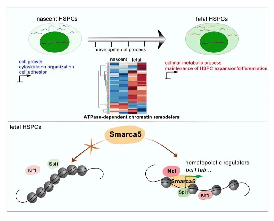
2020年8月5日,中国科学院动物研究所刘峰研究员团队与同济大学张勇教授团队合作在Blood杂志发表了题为Smarca5 mediated epigenetic programming facilitates fetal hematopoietic stem and progenitor cell development in vertebrates的论文。该工作发现:在产生和扩增/分化等不同发育阶段的造血干祖细胞中,染色质可接近性和转录组存在动态变化。深入机制探索发现,染色质重塑因子Smarca5通过与核仁蛋白Nucleolin相互作用,促进染色质重塑,调控造血相关的转录因子与基因组结合,进而促进造血干祖细胞发育。
为了寻找调控造血干祖细胞发育过程中染色质状态变化的关键染色质重塑因子,研究人员结合染色质重塑因子在AGM区和CHT区造血干祖细胞中的表达以及基因敲低/敲除实验,证实Smarca5通过调节染色质可接近性促进CHT区造血干祖细胞的增殖和分化。此外,通过质谱检测和功能性实验验证,研究人员发现核仁蛋白Nucleolin可以与Smarca5相互作用,并调控CHT区造血干祖细胞扩增/分化。进一步机制研究证明,Nucleolin有助于Smarca5介导的染色质重塑,进而影响与造血干祖细胞增殖和分化相关的转录因子(包括:Klf1和Spi1)与基因组的结合。例如:Smarca5缺失影响了Spi1在bcl11ab启动子区的结合,导致bcl11ab表达下调, 进而造成CHT区造血干祖细胞发育缺陷。
综上所述,该研究揭示了表观遗传调控在各造血组织中不同阶段的造血干祖细胞发育的重要性。
参考文献:
1. Laurenti E, Gottgens B. From haematopoietic stem cells to complex differentiation landscapes. Nature. 2018; 553(7689):418-426.
2. Orkin SH, Zon LI. Hematopoiesis: an evolving paradigm for stem cell biology. Cell. 2008; 132(4):631-44.
3. Zhang YF, Gao S, Xia J, et al. Hematopoietic Hierarchy - An Updated Roadmap. Trends in Cell Biology. 2018; 28(12):976-986.
4. Bertrand JY, Chi NC, Santoso B, et al. Haematopoietic stem cells derive directly from aortic endothelium during development. Nature. 2010; 464(7285):108-U120.
5. Kissa K, Herbomel P. Blood stem cells emerge from aortic endothelium by a novel type of cell transition. Nature. 2010; 464(7285):112-U125.
6. Boisset JC, van Cappellen W, Andrieu-Soler C, et al. In vivo imaging of haematopoietic cells emerging from the mouse aortic endothelium. Nature. 2010; 464(7285):116-U131.
7. Murayama E, Kissa K, Zapata A, et al. Tracing hematopoietic precursor migration to successive hematopoietic organs during zebrafish development. Immunity. 2006; 25(6):963-75.
8. Xue YY, Lv JH, Zhang CX, et al. The Vascular Niche Regulates Hematopoietic Stem and Progenitor Cell Lodgment and Expansion via klf6a-ccl25b. Dev Cell. 2017; 42(4):349-+.
9. McKinney-Freeman S, Cahan P, Li H, et al. The Transcriptional Landscape of Hematopoietic Stem Cell Ontogeny. Cell Stem Cell. 2012; 11(5):701-714.
10. Rybtsov S, Ivanovs A, Zhao SL, et al. Concealed expansion of immature precursors underpins acute burst of adult HSC activity in foetal liver. Development. 2016; 143(8):1284-1289.
11. Arora N, Wenzel PL, McKinney-Freeman SL, et al. Effect of Developmental Stage of HSC and Recipient on Transplant Outcomes. Dev Cell. 2014; 29(5):621-628.